Colorectal Quiz: May 8, 2023
Quiz for May 1:
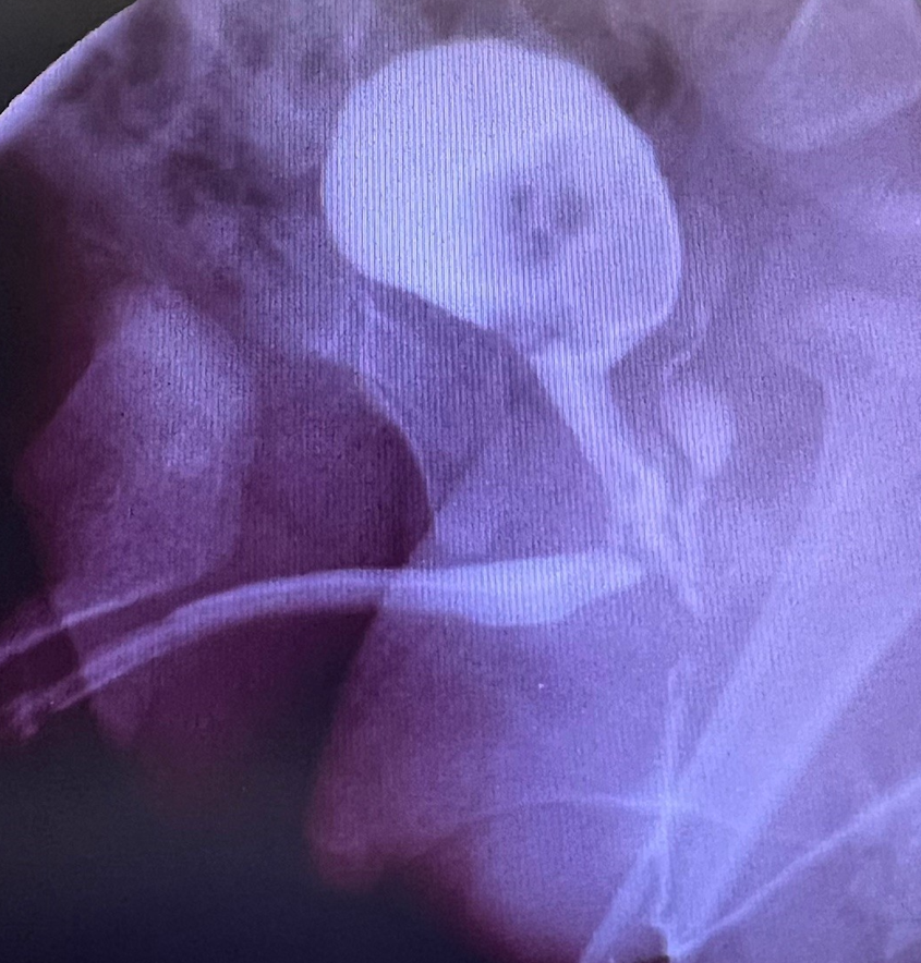
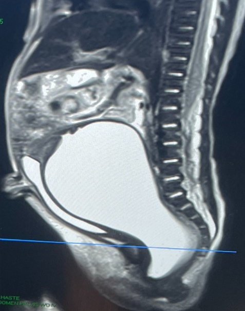
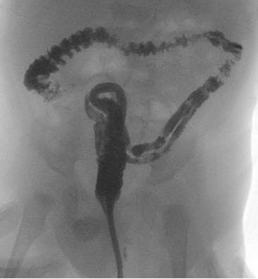
A newborn underwent fetal repair of a myelomeningocele. At birth, she was stooling and voiding spontaneously. But to ensure good bladder emptying was started on intermittent catheterizations. A routine VCUG was done and here is the image:
What do you think is going on? What would you do?
Answer for May 1st: This appears to be a colonic duplication with one of the rectums entering the urinary tract that other rectum exiting a normal anus. The rectum entering the urinary tract needs to be removed, with the good rectum left alone. This could be accomplished laparoscopically or via a combination of laparoscopy with a posterior sagittal approach. There appears to be a single lumen up to the splenic flexure, to the duplicated sigmoid can be left alone. If the entire colon were duplicated I would recommend mating the right colon’s two lumens anticipating that the patient might need antegrade flushes in the future.
Quiz for May 8:
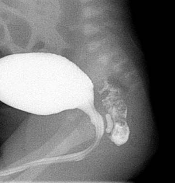
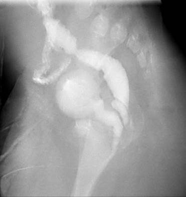
Here is the distal colostogram of a male ARM who underwent a colostomy at birth. It appears to show a bulbar fistula. But what is curious is the section just distal to the mucous fistula that appears narrow. How would you handle this case? PSARP and deal with the narrow area at the time of the colostomy closure? Resect the distal segment and pull through the proximal colostomy?
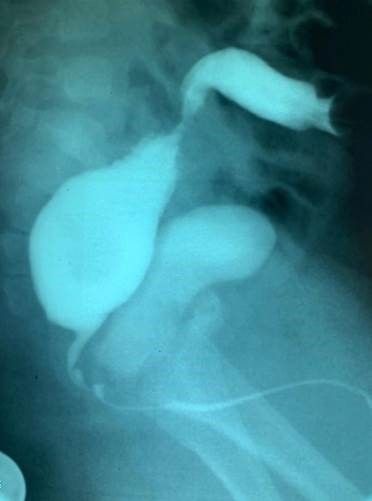
How would you handle this case?