Colorectal Quiz: June 12, 2023

Ron Reeder • June 12, 2023

Quiz for June 5:

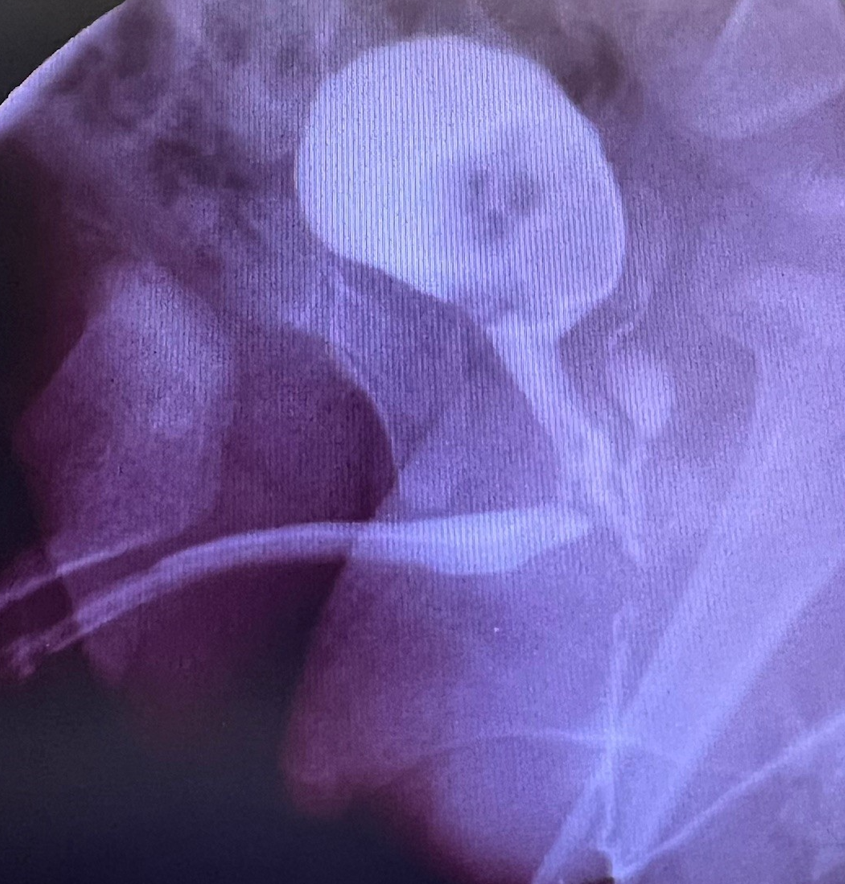
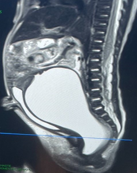
Baby girl, 8 months of age, with a urogenital sinus, and normal anus, without virilization. The imaging is below, contrast study and MRI. The blue arrow shows the vaginal to common channel fistula. Cystoscopy showed a common channel of 2 cm and a urethra length from fistula to bladder neck of 1 cm.

I asked how would you proceed?

Answer for June 5th Quiz

This patient with a UG sinus has a relatively low confluence but with a short urethra – a rare combination. If you did a total urogenital mobilization you would leave the patient with a too short urethra and they would leak urine. Then the option is to do a UG separation. Approaching this from above (laparoscopically or robotically) would be difficult to get to the vaginal-common channel fistula. Approaching that area perineally will also be difficult as there would be inadequate exposure. I would suggest a posterior sagittal approach with an ASTRA (cutting of the anterior rectal wall) or a full transanorectal approach (splitting the rectal wall in the anterior and posterior midline) to gain access. Then with that visualization you could mobilize up the anterior vaginal wall and close the common channel so the urethra becomes a combination of the common channel (2 cm) and the native urethra (1 cm). Then the vagina is pulled through and the rectum closed. It is safe to divert such a case.


Quiz for June 12:

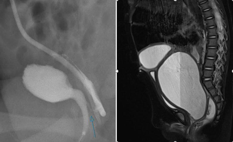
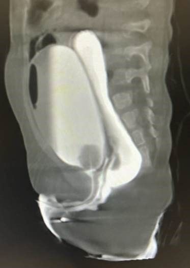
Here is the cloacagram from an 8 month old baby girl. She had a tethered cord which was detethered. What would be your approach? Posterior sagittal only? Total urogenital mobilization? Urogenital separation? How long would you leave the foley in? Would you use a suprapubic tube?

How would you handle this case?

By Ron Reeder January 29, 2024
Colorectal Conundrum for January 29: A male with a rectoperineal fistula undergoes a PSARP with mobilization of the rectum, both anterior and posterior rectal walls. In the days following surgery he starts to drain urine around the anoplasty consistent with a urethral injury. He is also voiding via the penis. A cystogram is shown below. How would you manage this situation? Answer: This patient has suffered from a urethral injury, and urine is draining out the posterior urethra into the perineum around the anoplasty. The key first step is to divert the urine with a suprapubic tube. A colostomy is not necessarily needed. The fistula might heal with diversion. If after a month or so a cystogram shows the persistence of the fistula, then a redo is needed with re-mobilization of the rectum, fully lifting the anterior rectal wall off of the urinary tract, and repair the urethra, with coverage of the posterior urethra with an ischiorectal fat pad. Then a voiding trial a month later with ultimate removal of the SP tube.
By Ron Reeder January 22, 2024
Colorectal Conundrum for January 22nd: In the previous week’s case, of an imperforate hymen, the MRI showed dilation all the way down to the perineum. Management involved a perineal – introital – incision to drain the fluid. If that were not the case and you had a dilated vagina but the distal extent was far away from the introital area, how might you handle that hydrocolpos? Answer: If this were a case of hydrolpos with normal anus and normal urethra, and the vagina cannot be drained by the introitus like an imperforate hymen, then drainage needs to be from above. IR or laparoscopy is an ideal approach to get a drain in the dilated structure (remember this could be bilateral, so both sides may need to be drained. Drainage is to relieve distension and most concerning its potential cause of distal ureteral obstruction causing hydronephrosis. Once the material hormones have dissipated the hydrocolpos will resolve and can be dealt with later in life via a laparoscopic distal vaginal pull-through, after the onset of puberty. In such a patient, I would follow closely with ultrasound 6 months after thelarche (breast budding).
By Ron Reeder January 16, 2024
Colorectal Conundrum for January 16: A newborn female is noted to have abdominal distension. On exam the uretha and anus are normal, and there appears to be a bulge in the hymen. The has an MRI with the image shown below. What would be your treatment plan? What are the variations in such a case and how would you manage each? Answer: This could be a case of imperforate hymen, but the MRI is key – how high up is the obstruction? How large is the hydrocolpos? In this case, it appears that a perineal incision might all that is needed as the dilation extends all the way down to the perineum. That is the most common scenario. An incision at the level of the hymen should drain out the fluid and no further intervention should be needed. Of course a careful exam needs to check to be certain you see a urethra – sometimes such a bulge could be a paraurethral cyst. For the next week’s question – what would you do if the perineal approach was inadequate – i.e. could not reach the dilated lumen.
More Posts
Share by: